Figure 6.

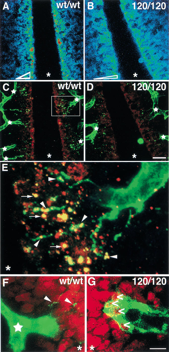
Filopodia extension toward VEGF-A deposits is impaired in the absence of heparin-binding isoforms. (A–D) Distribution of extracellular VEGF-A protein near the midline region of an 11.25-dpc wild type (A,C) and 120/120 (B,D) hindbrain. (A,B) Highest VEGF-A staining intensities are shown in red, lowest in blue; the shape of the VEGF-A gradient is shown at the bottom of the two panels. (C,D) PECAM-positive microvessels (green) extend many new branches (junctions labeled by white stars) toward the midline region in wt/wt (C), but not in 120/120 (D) hindbrains, in which, instead, the vessel network often terminates in closed loops. The microvessels closest to the midline extend multiple filopodia toward extracellular VEGF-A patches (red) in wt/wt hindbrains (C), but make only few filopodia in 120/120 hindbrains (D). (E) The area boxed in C, magnified 5×. A microvessel sprout extends filopodia (green) to contact VEGF-A patches (red); regions of overlap are indicated in yellow. (F,G) PECAM-positive vessel tips (green) near the midline region in wt/wt (F) and 120/120 (G) hindbrains; BrdU-positive (red) proliferating neuronal nuclei (in F,G) or double-positive endothelial nuclei (in G; open arrows). Some filopodia extending from the wild-type tip cell are indicated with arrowheads (F). The midline position in all panels is labeled with an asterisk. Bars: A–D, 20 μm; F,G, 100 μm.
