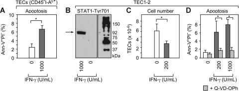
Figure 2.
IFN-γ activates STAT-1 in TECs and induces caspase-mediated programmed cell death. (A) Freshly isolated CD45−I-Ab int+high adult TECs (Figure S1) were cultured without (□) or with rmIFN-γ (▩) at the indicated concentration (U/mL). Apoptotic TECs (ie, Ann-V+PI−) were quantified after 72 hours of culture. A total of 4 experiments were performed. Bars depict mean ± SD; *P < .05. (B) TEC1-2 cells37 were cultured for 72 hours with rmIFN-γ and Tyr701 phosphorylated STAT-1 was detected by immunoblot analysis (∼92 kDa). (C-D) TEC1-2 were cultured with rmIFN-γ as indicated without or with (▨) the pan-caspase inhibitor Q-VD-OPh. Total cell numbers and apoptotic TEC1-2 were calculated from pooled data from 4 experiments. Bars depict mean ± SD; *P < .05.

