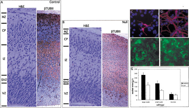
Figure 7.
N-Myc inhibits neuronal differentiation. (A,B) H&E (left) and βTubulinIII (right) staining of the control and null E17.5 cerebral cortex in the lateral ventricle region. Cells positively stained for β-tubulinIII are brown. (C,D) Anti-βTubulinIII immunofluorescence of control and null E12.5 neurosphere cultures stimulated to differentiate for 5 d. (E,F) Anti-GFAP immunofluorescence of control and null E12.5 neurosphere cultures stimulated to differentiate for 5 d. (G) Nuclear size in microns of the same neurosphere cultures as above. VZ, ventricular zone; SVZ, subventricular zone; IZ, intermediate zone; CP, cortical plate; MZ, marginal zone.

