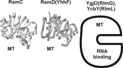
Figure 2.
The structure of ribosomal guanine-(N2)-methyltransferases. The structure of Mj0882 protein (12), which is likely to represent an ortholog of E. coli RsmC is marked as ‘RsmC’. The structure of E. coli RsmD(YhhF) protein is marked accordingly. Both protein structures are also marked ‘MT’ to indicate that they consist of single methyltransferase domain. For RlmG(YgjO) and RlmL(YcbY) the scheme is shown, where the protein is divided into N-terminal RNA-binding domain and C-terminal methyltransferase (MT) domain, based on conserved domain analysis (38).

