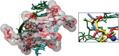
Figure 5.
Molecular (water)-accessible surface for the bound MTA molecule in the minor groove of a B-DNA of d(TCGCGA)2 sequence. Structural coordinates were taken from an NMR complex (16), PDB entry 146D. The figure was generated with the Chimera program (43). The right panel shows a detailed view of the region selected on the left panel. It also includes the structurally equivalent MSK atoms. This view highlights the different side-chains bound to the aglycone C-3 atoms in MTA (carbon atoms in blue) and MSK (carbon atoms in yellow). Compared to MTA, the shorter length of the MSK side chain restrains its possibilities of hydrogen bonding to DNA.

