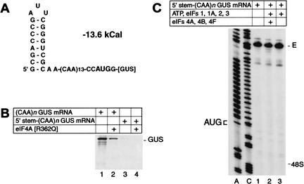
Figure 2.
Translation initiation on 5′-stem-(CAA)n-GUS mRNA. (A) Secondary structure of the 5′-UTR of 5′-stem-(CAA)n-GUS mRNA as predicted using mfold version 3.0 (Mathews et al. 1999). The theoretical standard free energy of the secondary structure is indicated. (B) Translation of (CAA)n-GUS and 5′-stem-(CAA)n-GUS mRNAs (0.2 μg) in RRL (15 μL) that had been preincubated without added mutant eIF4A (lanes 1,3) or with 1 μg R362Q mutant eIF4A (lanes 2,4) under conditions as described in the legend to Figure 1A. (C) Toeprint analysis of 48S complex formation on 5′-stem-(CAA)n-GUS mRNA in reaction mixtures that contained 40S subunits, GMP-PNP, and aminoacylated total tRNA in addition to translation components as indicated. Full-length cDNA is labeled E. The label “48S” indicates the expected position of toeprints caused by assembled 48S complexes on this mRNA. The position of the initiation codon is shown to the left of the two reference lanes, which show 5′-stem-(CAA)n-GUS sequence derived using the same primer as for toeprinting.

