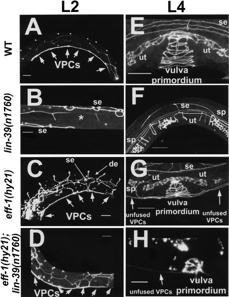
Figure 2.
eff-1 is epistatic to lin-39 during early and late cell-fusion events. Confocal reconstructions of worms carrying an ajm-1∷GFP construct marking the adherens junctions of epithelial cells (Shemer et al. 2000; Koeppen et al. 2001). The worms were assayed at early L2 (A–D) after the early fusion events of mid-L1 and at mid-L4 (E–H) after the late fusion events of mid-L3 (see Fig. 1 for details). (A) Wild-type worms showing the six VPCs that escape fusion to the hypodermis after they attach to each other (n > 100). (E) Three of these VPCs escape cell fusion to hyp7 at L3, and their 22 great-granddaughters invaginate, forming a stack of seven rings (n > 100). (B,F) In lin-39(n1760) single null mutants, no VPCs formed after all of the Pn.p cells had fused to the hypodermis (*, n = 50), resulting in the absence of a vulva (Vul phenotype) at late L4 (F; n = 100). (C) eff-1(hy21) mutants grown at the restrictive temperature of 25°C showing the VPCs that fail to fuse with hyp7 (n = 50). P3.p, P4.p, and P8.p also fail to fuse in the L3 and attach to the vulva primordium formed from the descendants of P(5–7).p. (G) The result is a stack of rings connected to a row of ectopic cells (n = 70). Due to fusion failure in eff-1(hy21), ectopic dorsal epithelia (de) and lateral hypodermal seam cells (se) are present and migrate throughout the body of the worm. (D,H) Pn.p cells fail to fuse in eff-1(hy21);lin-39(n1760) double mutants at L1 (D; n = 45) and later at L3 (H; n = 28). The unfused VPCs migrate and invaginate, forming a “pseudo” vulval primordium that is incomplete and abnormal structurally, resulting in a nonfunctional vulva (cf. H and G,E). ut, uterus; sp, spermatheca. Anterior is to the left and dorsal is up, except for B, a ventral view. The fusion status of the cells was also confirmed by staining worms with the MH27 antibody (Podbilewicz and White 1994). Arrows mark unfused cells. Bar, 10μm.

