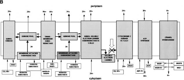
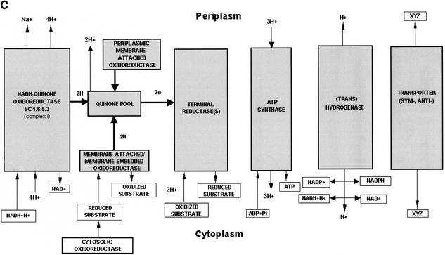
Figure 3.
Graphical visualization of the functional reconstruction of Xylella fastidiosa aerobic and anaerobic respiration. (A) Diagrammatic representation of aerobic respiration. Three-dimensional boxes show the presence of functional components and filled arrows indicate electron flow. (B) Detailed spatial disposition of membrane components of aerobic respiratory chain. The absence of cytochrome c oxidase is designated by the cross and arrow. (C) Overview of the anaerobic respiratory chain. Reconstructions show proton translocation and oxidative phosphorylation.


