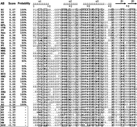
Figure 2.
Multiple alignment of the BirA N-terminal domains and identification of the HTH motif. The known secondary structure of the Escherichia coli BirA is shown in the first row. The α2 and α3 helices form the helix–turn–helix (HTH) structure. The score and the probability of the candidate HTH motif are given. A score of <2.5 is not significant. Non-HTH proteins are boxed, except BirA from Bacillus cereus, which is a false-negative prediction (see text). The genome abbreviations are listed in Table 1.

