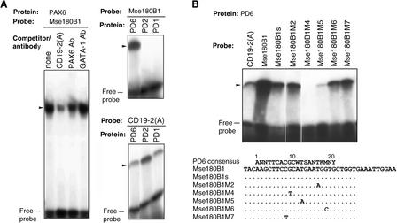
Figure 1.
(A)EMSAs of the PD6 binding site Mse180B1. CD19-2(A) was used as a positive control. PAX6 binding competitor CD19-2(A) and rabbit serum (PAX6 and GATA-1) were added to the EMSA reactions. (B) EMSA of Mse180B1 mutants and the probe sequences, aligned with the PAX6 consensus binding sequence; (W) A or T, (S) C or G, (K) G or T, (M) A or C, (R) A or G, (Y) T or C, and (N) G, T, A, or C. Dots represent nucleotides identical to those in Mse180B1. The proteins and peptides used for binding were in vitro synthesized mouse PD1 and PD2, human PD6, the N-terminal half of PAX6, including the PD and HD domains (PD + HD), and the entire PAX6 protein (PAX6).

