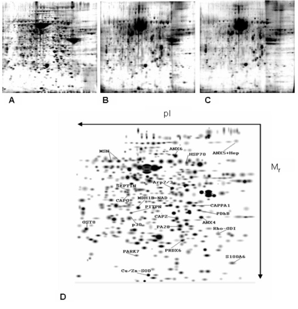
Figure 2.
Protein maps representative of each of the treatment sub-groups analyzed in side by side comparison and filtered baseline reference map annotation. A. Control treatment, B. heparin 24 hours, C. heparin 96 hours, and D. a computer analyzed, 2D Gaussian filtered lung reference map with the identified protein spots, separated based on pI and molecular weight (Mr). Note repeatability between sub-groups.

