
Figure 1.
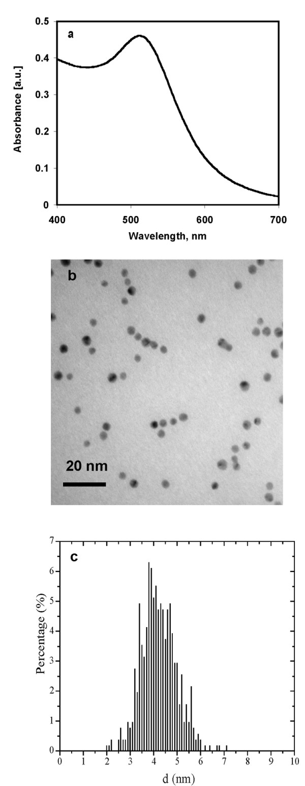
UV-Visible spectroscopy and transmission electron micrograph of gold nanoparticles. a) UV-visible spectrum of a gold nanoparticles solution and b) Transmission electron microscopy picture of gold nanoparticles. TEM was done after drop coating the gold nanoparticles on carbon coated copper grid, c) histogram showing the size distribution of gold nanoparticles.