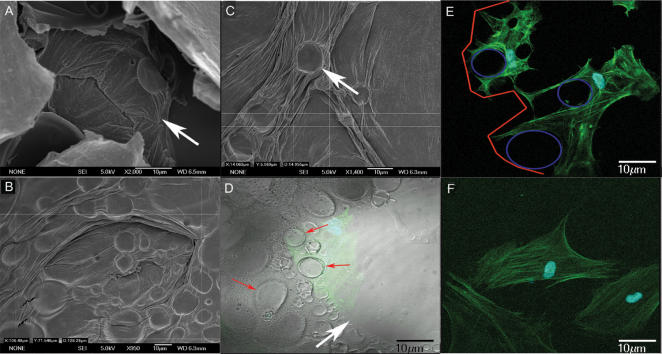
Figure 1. Images showing primary and secondary micro concavities at scanning electron and confocal microscopy.
(A) Primary micro concavity (arrow) of the PLGA surface at SEM. Cells can be completely contained within a primary concavity, due to its dimensions. (Calibration Bar = 10 µm); (B) SEM analysis of primary concavity dimensions (Calibration Bar = 10 µm); (C) SEM analysis of secondary concavity dimensions (Calibration Bar = 10 µm); (D) The interaction between the concave surface, showing primary (white arrow) and secondary (red arrows) micro-concavities at the confocal microscope (in green a cell within a concavity). The intimate adherence of a cell to the polymer surface and its nuclear polarity are clearly observable. The image was been obtained superimposing dark field with light field confocal microscopy (Calibration Bar = 10 µm); (E) Confocal image showing primary (outlined in red) and secondary (outlined in blue) micro-concavities and spider-shaped cellular elongations (Calibration Bar = 10 µm); (F) A gingival fibroblast not showing cellular alterations or nuclear polarity at the confocal microscope (Calibration Bar = 10 µm).

