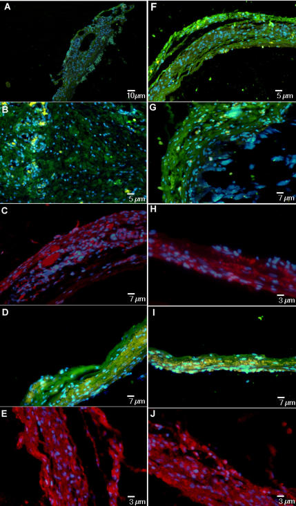
Figure 7. Immunofluorescence confirming the presence of a mineralized extra cellular matrix on concave texturing.
The panel shows positivity for Collagen I (A) FITC (green) (Calibration Bar = 10 µm), BAP [Bone Alkaline Phosphatase] (B) FITC (green) (Calibration Bar = 5 µm), OC [Osteocalcin] (C) PE (red) (Calibration Bar = 7 µm), ON [Osteonectin] (D) FITC(green) (Calibration Bar = 7 µm) and BSP [Bone Sialoprotein] (E) PE (red) (Calibration Bar = 3 µm). The same analysis confirming the presence of a mineralized extra cellular matrix on smooth texturing. The panel shows positivity for Collagen I (F) FITC (green) (Calibration Bar = 5 µm), BAP (G) FITC (green) (Calibration Bar = 7 µm), OC (H) PE (red) (Calibration Bar = 3 µm), ON (I) FITC (green) (Calibration Bar = 7 µm) and BSP (J) PE (red) (Calibration Bar = 3 µm). Nuclear staining is obtained with DAPI (blue).

