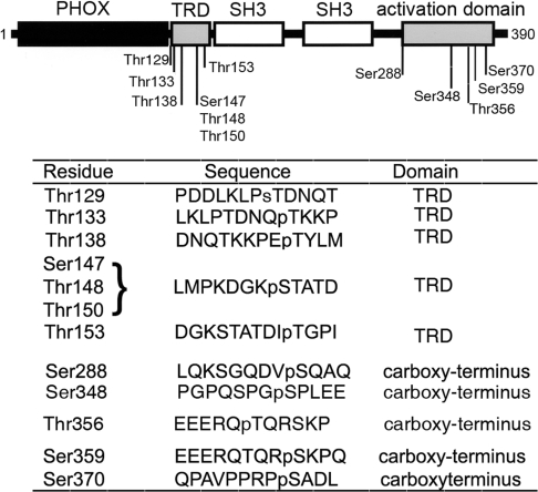
Figure 2. IRAK-4 dependent phosphorylation sites identified in p47phox by microcapillary HPLC–MS/MS.
IRAK-4-dependent phosphorylated sites in p47phox were detected using microcapillary reversephase HPLC–nanoelectrospray and targeted ion MS/MS as described in the Experimental section. The upper panel shows a schematic representation of p47phox. The phox homology (PHOX) domain is shown in black, the SH3 domains are shown in white, and the phosphorylation domains are in grey. The lower panel shows the sequences surrounding the phosphorylated residues detected by HPLC–MS/MS. Phosphorylated residues are preceded by ‘p’. Owing to the proximity of Ser147, Thr148 and Thr150, the precise phosphorylation site could not be resolved, although a definitive phosphorylation site exists at these residues. For the complete MS/MS spectra, see Supplementary Figure S1 at http://www.BiochemJ.org/bj/403/bj4030451add.htm.

