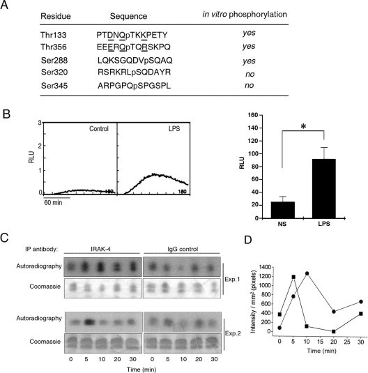
Figure 7. Identification of phosphorylated residues in endogenous p47phox following LPS stimulation.
(A) The phosphorylated sites in endogenous p47phox immunoprecipitated from LPS-stimulated neutrophils were detected using microcapillary reverse-phase HPLC–nanoelectrospray and targeted ion MS/MS as described in the Experimental section. The sequences surrounding the phosphorylated residues detected by HPLC–MS/MS in endogenous p47phox are shown. Phosphorylated residues are preceded by ‘p’. (B) ROS production in human neutrophils after LPS stimulation was measured by luminol-dependent chemiluminescence. Kinetics representative of three different experiments (left-hand panel) and means±S.E.M. for three different experiments (right-hand panel) are shown. RLU, relative light units. (C and D) IRAK-4 activity in human neutrophils after LPS stimulation. IRAK-4 was immunoprecipitated (IP) from lysates obtained from neutrophils after stimulation with LPS. IRAK-4 activity was determined by the phosphorylation of the substrate histone H1.2 in the presence of 4 μCi of [γ-32P]ATP. Phosphorylation was analysed by autoradiography. (C) Results are from two different experiments. (D) The signal of phosphorylation for each time point was quantified using Quantity One 4.2.1 software. The background for each time point (IgG control) was subtracted from the IRAK-4 signal. The kinetics for two independent experiments are shown: ●, experiment 1; ■, experiment 2.

