
Figure 3.
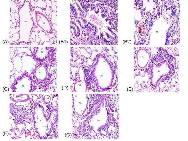
Effects of ketamine on histopathological changes seen in pulmonary sections from ovalbumin-sensitized and -challenged rats. Representative paraffin-embedded, Hematoxylin and Eosin-stained lung sections were prepared from the left lungs of experimental rats, showing bronchioles, lung alveoli and surrounding vessel structures. The groups are as shown in Figure 1. (A) PBS; (B1-B2) OVA; (C) 12.5 mg/ml; (D) 25 mg/ml; (E) 50 mg/ml; (F) 50 μg/kg; (G) 100 μg/kg. VL, vascular lumen; AL, airway lumen. Arrows demonstrate marked perivascular edema (B1, G), places where inflammation has destroyed a portion of the airway epithelium (B2, F), and eosinophils (B1, C, E). Arrowheads indicate peribronchial inflammation (B1, C, D, E, F, G), hemorrhage (B2, E, F, G) and congestion (B2). Sections were evaluated under light microscopy. Original magnification was ×40, and the scal bars represent 20 μM.