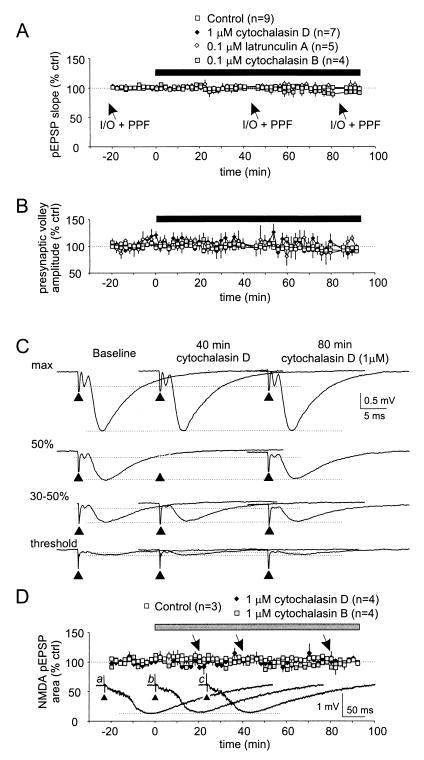
Figure 1.
Actin assembly inhibitors do not affect basal synaptic transmission. (A–C) Bath-applied cytochalasin D, cytochalasin B, and latrunculin A affect neither the pEPSP slope nor the presynaptic fiber volley amplitude. After establishing baseline recordings, cytochalasin D (1 μM, n = 4; 0.1 μM, n = 3; data pooled), cytochalasin B, or latrunculin A was continuously superfused (solid bar). Stimulation intensities were set to evoke 30–50% of maximal pEPSP amplitude. Data gaps at 40 and 80 min are because of additional I-O protocols. (A) Although the average pEPSP slope declined slightly at 80 min after cytochalasin B, none of the tested AAIs induced significant changes as measured after 20, 40, 60, and 80 min. (B) Parallel analysis of the corresponding fiber volley amplitudes showed no detectable changes. (C) Traces (averages of two) from a series of I-O curves taken under baseline, 40, and 80 min after the start of cytochalasin D superfusion. (D) NMDA–pEPSPs were isolated as described in Methods and cytochalasin D or cytochalasin B at 1 μM continuously superfused (gray bar). Neither the NMDA–pEPSP nor the corresponding presynaptic volley was significantly affected. Even after 80 min of cytochalasin treatment, the area of the NMDA–pEPSP did not vary significantly from control. Inset shows representative raw traces taken at 10 min before (a), 40 (b), and 80 (c) min (arrows) after the start of superfusing cyotchalasins. Arrowheads mark the stimulation artifacts (truncated).

