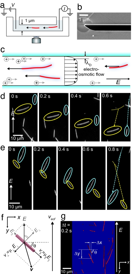
Fig. 2.
Experimental geometry. (a) Electrophoretic motion of microtubules in channels is observed using fluorescence microscopy. (b) Scanning-electron microscope image of a part of the cross-section of a channel. (c) The velocity of microtubules is a superposition of their electrophoretic velocity and an EOF velocity. (d and e) Fluorescence of electrophoresis (E = 4 kV/m) of individual microtubules. (d) A microtubule oriented axially along the electric field moves faster than a microtubule oriented perpendicular to the field. (e) Microtubules can have a velocity that is not collinear with E. (f) Diagram of the velocity components of a microtubule oriented under an angle θ with E along the y axis. The electric field has components axially (E‖) and perpendicular (E⊥) to the microtubule. The anisotropic mobility results in net velocity v that is not collinear with E. (g) Overlay of two images taken at a 0.2-s interval. For each microtubule, the average orientation θ and center-of-mass displacements in the x and y directions, Δx and Δy respectively, are determined.

