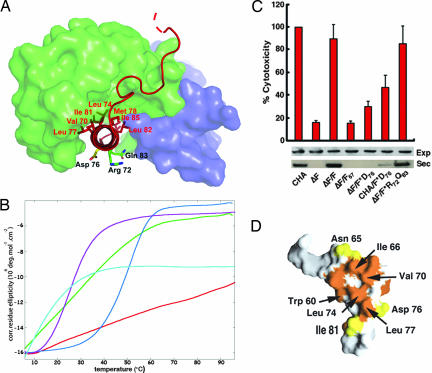
Fig. 3.
PscG grasps PscF's amphipathic tail. (A) Hydrophobic residues within the C-terminal helix of PscF (PscF68–85, in red) are shielded within PscG's concave groove, whereas hydrophilic residues point to solvent. PscG and PscE are represented as light green and blue surfaces, respectively. (B) Thermal denaturation curves obtained by circular dichroism measurements of wild-type PscF (red), PscF V70K/I77K (green), PscF L74K/I81K (purple), PscF M78K/L82K (blue), and PscF1–67 (light blue). Wild-type PscF is the only molecule that is robustly resistant to thermal denaturation. (C) Cytotoxicity profiles of P. aeruginosa strains carrying a pscF chromosomal deletion complemented by either wild-type (ΔF/F) or mutant pscF-expressing plasmids. Strains complemented by wild-type PscF or PscF R72A/Q83A are as cytotoxic as wild-type (CHA), whereas the one complemented with PscF D76A displays greatly reduced cytotoxicity. Supplying the wild-type strain with PscF D76A generated a dominant-negative effect. Below, Western blots revealing the cytoplasmic expression (exp) and secretion (sec) of T3SS effector molecule PopB in the same strains. Although all strains express PopB in the cytoplasm, only strains carrying either wild-type PscF or PscF R72A/Q83A are able to secrete PopB at levels comparable to the wild-type (CHA) strain. Error bars refer to standard deviation measurements for experiments performed in triplicate. (D) Mapping of PscF residues that are identical (yellow) and similar (orange) within different bacterial species from the point of view of PscG's concave side.

