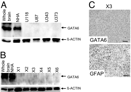
Fig. 3.
Alterations in GATA6 expression among human GBM lines and explant xenografts. (A) Western blot analysis demonstrating loss of GATA6 expression in all four established human GBM lines. Each lane has 40 μg of total protein lysate, with β-actin used as a positive loading control. Whole-brain sample was obtained from a head injury patient requiring surgery, whereas NHA represents human hTERT immortalized but nontransformed astrocytes. (B) GATA6 expression was absent in six human GBM explant xenografts. Each lane has 40 μg of total protein lysate, with β-actin used as a positive loading control. (C) GATA6 expression in human GBM explant xenograft (X3) determined by IHC. A rabbit GATA6 polyclonal primary antibody and a protein G-HRP secondary antibody were used. None of the xenografts expressed GATA6, with 50% immunopositive for the differentiated astrocyte marker GFAP (rabbit GFAP polyclonal antibody from DAKO). No immunoreactivity was detected on the GBM explant xenograft specimens using only a secondary antibody. (Magnification: ×200.)

