Fig. 5.
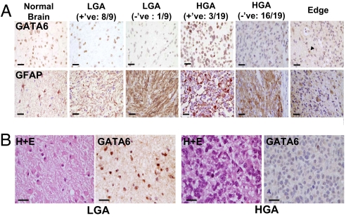
Screening of human astrocytomas operative specimens for alterations in expression using IHC. (A) GATA6 expression was tested by IHC in normal human brain (from head injury patient), nine LGA, and 19 human GBM paraffin-embedded specimens. One of nine (≈10%) LGA and 16/19 (≈85%) GBM had loss of GATA6 expression, suggesting that loss of GATA6 is involved in astrocytoma progression. Pictures show GATA6 (Upper) and GFAP (Lower) immunostaining. The rightmost panels demonstrate the invading edge of a GBM with loss of GATA6 expression, while nontransformed astrocytes and other cells retain GATA6 expression (arrowhead). (Magnification: ×400. Scale bars: 25 μm.) (B) GATA6 expression is analyzed in a patient with a pathologically documented secondary GBM. GATA6 expression is abundant in the initial resected LGA (Left) but absent when the patient recurred with a GBM (Right). Shown are H&E immunostaining and GATA6 immunostaining. (Magnification: ×400. Scale bars: 25 μm.)

