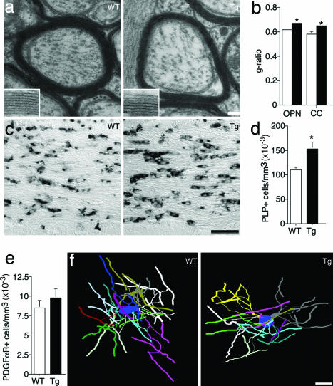
Fig. 2.
OL structure and number are altered in the absence of erbB signaling. (a) Representative EM of myelinated axons in the corpus callosum. (Insets) Higher magnification of myelin in WT and Tg animals. (b) Quantification of the g-ratio in WT (open bars) and Tg (filled bars) optic nerve (OPN) and corpus callosum (CC). Tg mice have thinner myelin sheaths than WTs (P = 0.031). (c) Representative images of PLP in situ hybridization in corpus callosum sections. (d) Quantification of PLP+ cells in corpus callosum shows a higher density of OLs in Tg mice (P = 0.007). (e) The density of PDGFαR+ OL progenitors is not altered in Tg mice (P = 0.36). (f) Three-dimensional reconstructions of representative WT and Tg frontal cortex OLs. For quantification of OL morphology see Table 1. Error bars represent SEM. (Scale bars: 80 nm in a, 26 nm in Insets, 50 μm in c, and 20 μm in f.)

