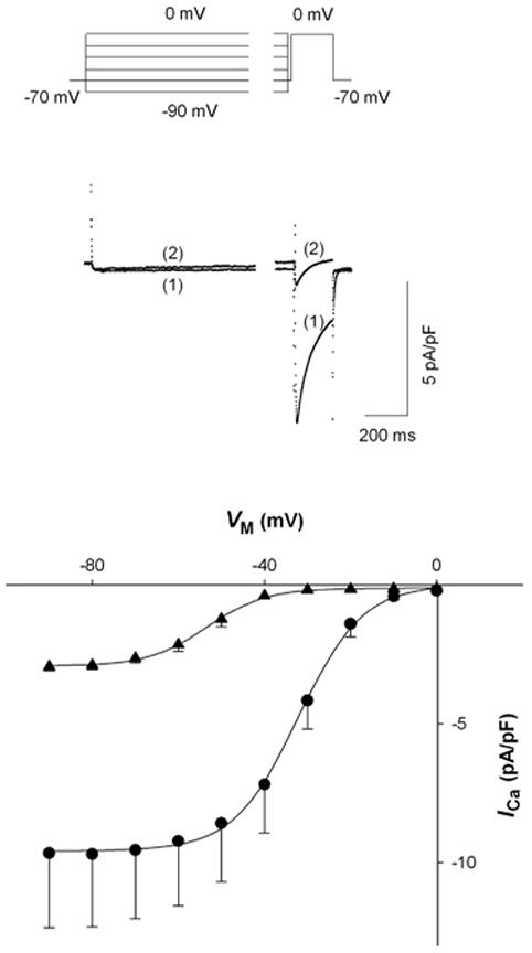
Figure 2.

Effect of [Mg2+]p on the VM dependence of steady state ICa inactivation. Two-pulse inactivation protocol with a 20 sec prepulse and 200 msec test depolarization to 0 mV (top). Typical currents with a prepulse of -30 mV where traces (1) and (2) were recorded with 0.2 and 1.8 mM [Mg2+]p, respectively (middle). ICa plotted versus prepulse potential (VM) for myocytes voltage-clamped at 0.2 (● , n=6) and 1.8 mM [Mg2+]p (▲ , n=6) (bottom).
