
Fig. 3.
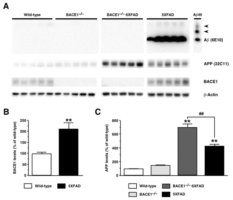
Effects of BACE1 null mutation on cerebral Aβ, BACE1 and full-length APP levels in 5XFAD transgenic mice. (A) Immunoblot analyses of protein extracts (15 μg/lane) from hemibrain homogenates of wild-type control, 5XFAD, BACE1−/−·5XFAD, and BACE1−/− mice at 18 months of age. Shown are immunoblots for Aβ (top panels; anti-total Aβ antibody 6E10), APP (second from top panels; anti-mouse and human APP antibody 22C11), BACE1 (third from top panels; anti-BACE1 antibody BACE1-Cat), and actin (bottom panels; anti-actin antibody AC-15). 5XFAD brain homogenates show a strong Aβ band, while Aβ is below the level of detection by immunoblot analysis in brain extracts from the other three genotypes. The lane labeled “Aβ40” contains 100 ng of synthetic Aβ40 for use as a positive control; the two slower migrating bands (arrowheads) likely represent multimeric Aβ assemblies. (B) Quantification of BACE1 blots in which band intensities were measured by phosphorimaging and expressed as percentage of wild-type control levels. Note that BACE1 levels are significantly elevated in 5XFAD brains. (C) Quantification of APP blots in which band intensities were measured by phosphorimaging and expressed as percentage of wild-type control levels. Note that while 5XFAD mice overexpress human APP approximately fourfold relative to endogenous mouse APP protein, levels of APP are elevated even further in BACE1−/−·5XFAD mice. Data are presented as the mean ± SEM of 5 animals. **P < 0.01 (vs. wild-type controls), ##P < 0.01 (vs. BACE1−/−·5XFAD).