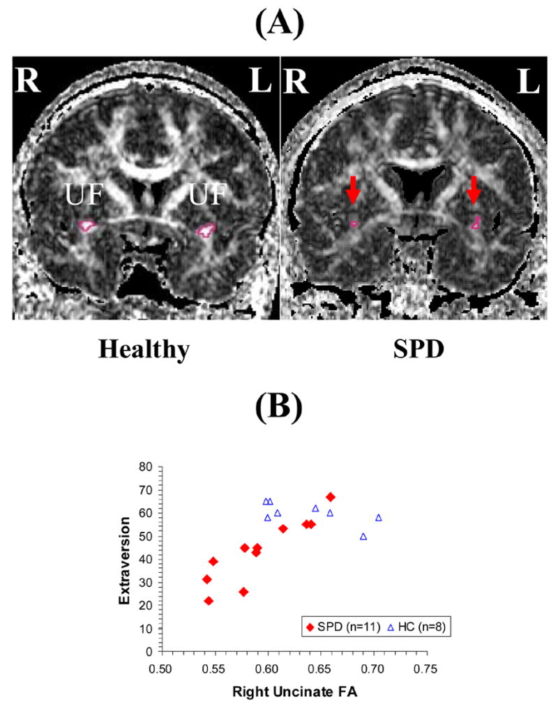
FIGURE 1. The Uncinate Fasciculus (UF) and Extraversion in SPD.

(A) Line-scan diffusion tensor imaging (LSDTI) produced these contrasting coronal views of uncinate fasciculi (UF) in comparison (left) and SPD (right) subjects.
(B) Graph illustrates the relationship of extraversion to right uncinate fasciculus (UF) fractional anisotropy (FA) in SPD (Pearson r= .89, p= .0002) and comparison (r= −.66, p= .075) subjects.
