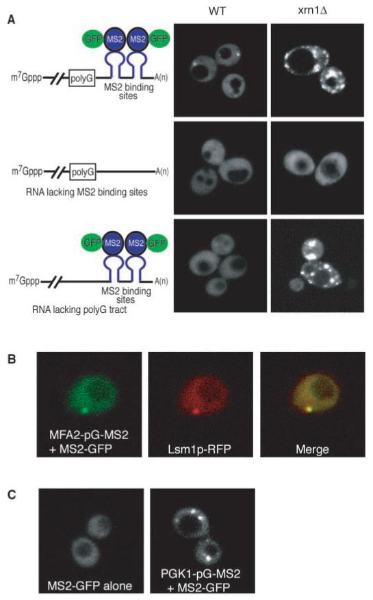
Fig. 3.

RNA decay intermediates are localized to P bodies. (A) WT (left column) and xrn1Δ (right column) cells are shown expressing the MFA2 mRNA with poly(G) and MS2 sites (top row), or the MFA2 mRNA with only a poly(G) tract (middle row), or the MFA2 mRNA with only the MS2 sites (bottom row). The schematic diagram of the reporter RNA expressed from the plasmid and its interaction with the MS2-GFP fusion protein is shown on the left. For simplicity, only a single protein is shown bound to a stem loop; however, because MS2 coat protein binds as a dimer, at least two MS2-GFP molecules are bound per site. (B) Left, RNA; middle, Lsm1p-RFP; and right, the merge generated by Adobe Photoshop. (C) WT cells expressing only the MS2-GFP fusion protein (left) or with a PGK1 reporter mRNA with a poly(G) tract and MS2 binding sites (right).
