
Figure 2.
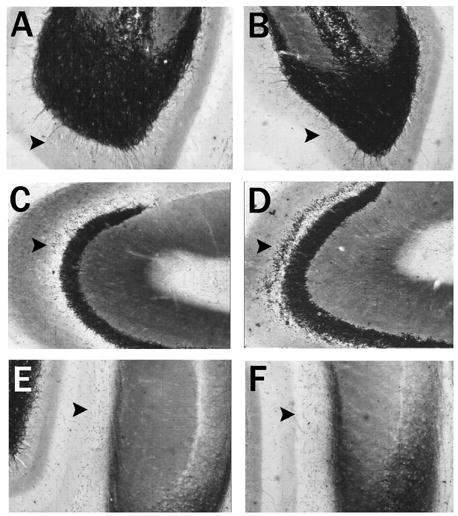
Hippocampus horizontal brain sections stained with Timm histochemistry after saline and kainic acid on postnatal day 15. Timm’s histochemistry depicts alterations in the laminar pattern of staining three weeks after rats experience an episode of convulsive status epilepticus induced by kainic acid (KA-SE) on postnatal day 15 as compared to controls. The inner molecular layer of the dentate gyrus (arrow) shows few or no punctate granules three weeks after saline (A) injections on postnatal day 15 (P15) or KA-SE on P15 (B). As compared to A, there are no alterations in the inner molecular layer of the dentate gyrus. The stratum oriens of CA3 in a normal control rat (C) sacrificed three weeks after saline injections on P15 shows few punctate granules (arrow). However, the stratum oriens of CA3 three weeks after a rat experienced KA-SE on P15 (D) developed a band of Timm granules in the stratum oriens of CA3 indicating sprouting of the mossy fiber projection to this region. These differences were quantified in Table 1. The stratum moleculare of the ventral CA1 region in a normal rat (E) sacrificed three weeks after saline injections on P15 shows few or no granules in this region (arrow); similar to rats that experienced KA-SE on P15 (F). As compared to E, there were no alterations in the pattern of Timm staining in this region.