
Fig. 3.
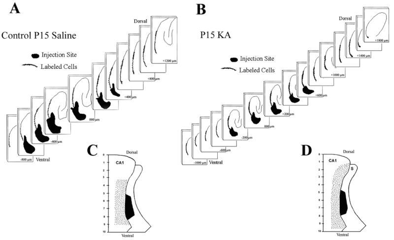
A. & B. Serial Neurolucida reconstructions of the rat hippocampus in horizontal brain sections of sodium selenite retrograde tracing in a rat that received saline injections (A) on postnatal day 15 (P15) and a rat that experienced convulsive status epilepticus induced by kainic acid (KA-SE) (B) on P15. The series of two-dimensional reconstructions demonstrate the dorsoventral extent of the injection site in the subiculum (dark filled area), and the larger dorsoventral extent of the retrograde labeling in the CA1 pyramidal region (dots depict labeled cells). Sections are arranged serially from the most dorsal section with labeling at the upper right corner to the most ventral section demonstrating labeling in the lower left corner. C. & D. Uni-dimensional flat maps of the dorsoventral extent of retrograde sodium selenite labeling in the CA1 pyramidal region and injection site in the subiculum. Saline treated controls (C) demonstrate a smaller dorsoventral extent of retrograde labeling in the CA1 pyramidal region as compared to a rat that experienced an KA-SE on P15 (D). The ratios of CA1 retrograde labeling / subiculum injection site for both groups of rats are shown in Table 2.