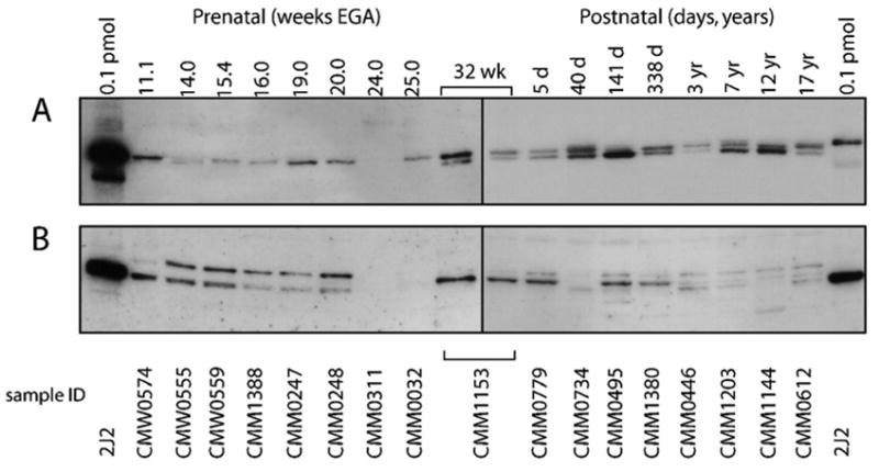
Figure 3. CYP2J2 immunoreactive protein in fetal and postnatal liver.

A representative set of samples ranging from 11.1 to 32 weeks EGA and 5 days to 17 years postnatal age are shown. Each lane contained 4 μg microsomal protein, the first and last lanes contained 0.1 pmol recombinant CYP2J2 protein as a positive control. Sample CMM1153, which exhibits a pattern that is more consistent with that observed in the postnatal samples, was loaded on both blots as a reference for band patterns. Membranes containing the fetal and postnatal samples were exposed for 30 and 10 seconds, respectively.
A: detection of immunoreactive protein with anti-CYP2J2rec
B: detection of immunoreactive protein with anti-CYP2J2pep3
