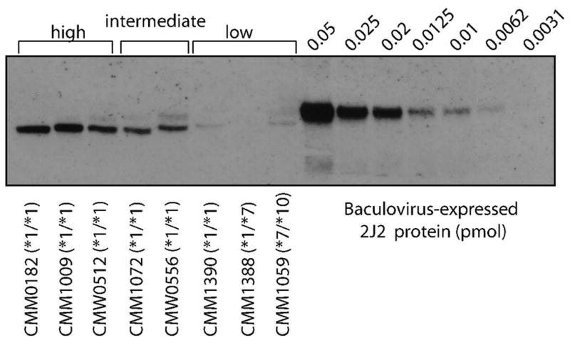
Figure 4. Variability of fetal CYP2J2 immunoreactive protein and relationship between protein content and mRNA expression.

Each lane contained 4 μg microsomal protein. Anti-CYP2J2 antibody was used for panels A and B.
Immunoblot with representative samples demonstrating the range of immunoreactive protein observed. For descriptive purposes, ‘low’, ‘intermediate’ and ‘high’ protein categories were assigned by visual inspection. There was no relationship between genotype and amount of protein observed.
