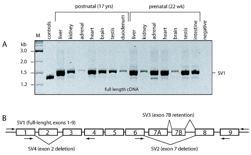
Figure 5. Expression of CYP2J2 splice variants.

Two sets of tissues, one from a postnatal (17 year old) and one from a fetal (22 wks estimated gestational age) male, were utilized for alternate splice variant analysis. Each reaction contained cDNA corresponding to 40 ng total RNA.
A: Full-length cDNA (SV1) was the major amplification product in each tissue analyzed. Minor variants were visible across the panels. M, 1 kb ladder (A); control, amplification from a plasmid mix containing the exon 2 and exon 7 deletions.
B: Graphic display of alternate splice variants identified by cloning and sequencing. Primer locations are as indicated. Boxed depict exons. Transcript-specific PCR for the exon 2 deletion variant (SV4) did not produce any visible product (not shown).
