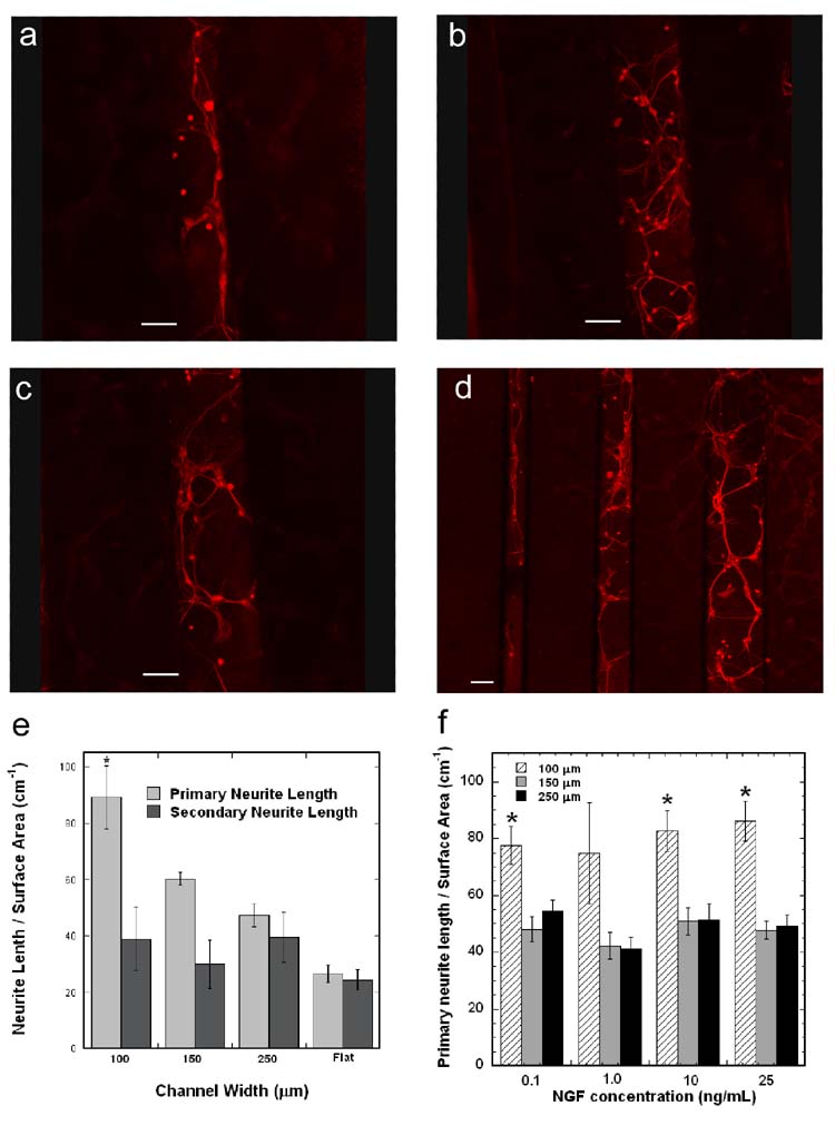
Figure 5. Neuronal co-cultures within PLG microchannels.
Fluorescence imaging of neurons cultured within PLG channels with immobilized pNGF/PEI complexes: 100 μm (a), 150 μm (b), 250 μm (c), and all channel widths (d). Scale bars represent 100 μm. Length of primary and secondary neurites per surface area within the PLG channels and on flat PLG disks with immobilized pNGF/PEI complexes (e). Length of primary neurites per surface area within PLG channels with various NGF concentrations added to the culture media (f). The symbol * indicates statistical significance, p < 0.05.

