
Figure 2. Failure of BrdU-positive cells to leave the ventricular surface and PVH in MEKK4-deficient forebrain.
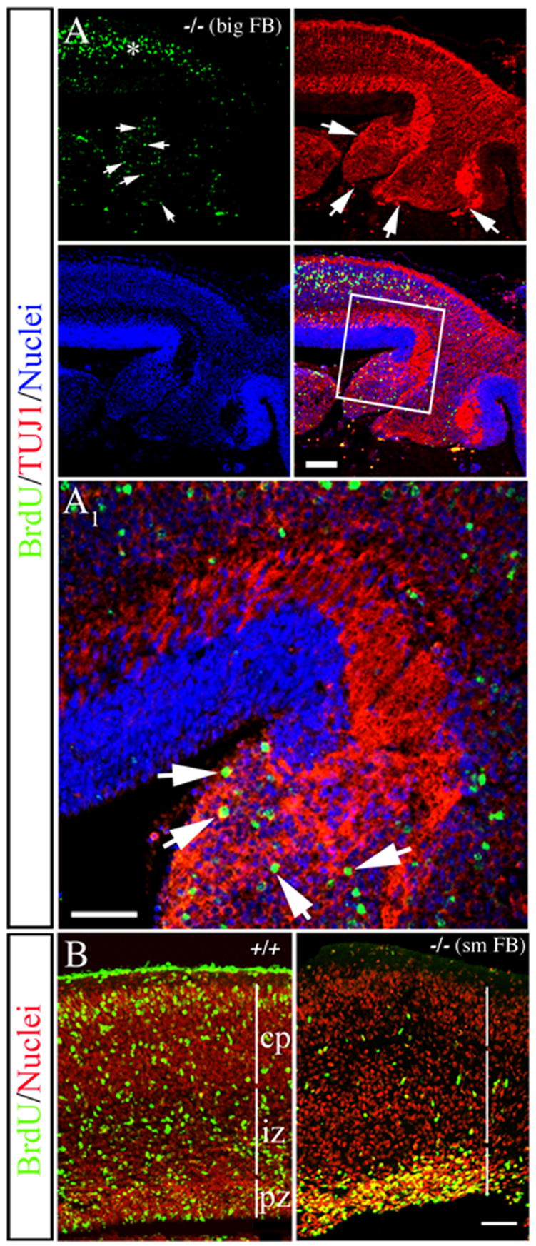
(A and B) Dams were exposed to BrdU at E14.5 and the forebrain (FB) was analyzed at E18.5.
(A) Analysis of a PVH from a MEKK4−/− “big FB” phenotype stained for BrdU (green), TUJ1 (red) and TO-PRO-3 (blue). Compared to adjacent neocortex (asterisk), heavily labeled BrdU+ cells (arrows in BrdU) co-localized with TUJ1+ (arrows) in the PVH. (A1) Higher magnification of the boxed region in A shows heavily labeled BrdU+/TUJ1+ cells (arrows) within the heterotopia. Bars = 50μm.
(B) Example of a MEKK4+/+ and MEKK4−/− with a “small FB” phenotype (sm FB) where BrdU+ cells (green) failed to leave the VZ surface compared to +/+. Nuclei were labeled with propidium iodide (red). Bar =100 μm.