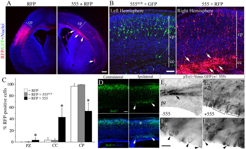
Figure 3. MEKK4 siRNA impairs radial migration of neocortical neurons.

(A, C, D and E) In utero electroporation was performed at E14.5 and forebrains were examined at P0.
(A) Transfection with RFP alone (left) resulted in the majority of cells reaching the cortical plate (CP). In contrast, RFP+ cells co-transfected with siRNA#555 (arrows, right) were largely stuck in the corpus callosum (CC). Bar = 200μm.
(B) Example of an E15.5 brain electroporated with siRNA#555SCR and GFP into the left hemisphere and siRNA#555 and RFP into the right hemisphere. At P0, siRNA#555 caused the majority of RFP+ cells to arrest in the CC whereas most GFP+ cells reached the CP. Bar= 50μm.
(C) Quantification of the percentage of RFP+ cells in the proliferative zone (PZ), CC or CP from mice electroporated at E14.5 and sacrificed at P0. siRNA#555 (black bars) caused ~50% reduction of cells in the CP and significantly more cells to accumulate in the CC and PZ compared to RFP alone (white bars) or siRNA#555SCR (grey bars). *p<0.01 (ANOVA).
(D) In utero electroporation of fetuses at E14.5, exposure of dams to BrdU at E15.5, and analysis at P0. Example of a brain electroporated with siRNA#555 shows a heterotopia (arrows) beneath the CP (ipsilateral) that contained many BrdU+ cells (arrows). In the ipsilateral area of CP (corresponding to the asterisk in lower panel) reduced BrdU immunostaining is observed over the heterotopia compared to the adjacent CP or opposite hemisphere (contralateral). Bar =100 μm.
(E) Electroporation of pTα1-Venus-GFP alone (E1) or with siRNA#555 (E2–4) and immunostaining for GFP. (E1) Electroporation of pTα1-VenusGFP alone (−555) resulted in most GFP+ cells reaching the cortical plane (not visible in picture) with their axonal projections (arrow). (E2) Co-electroporation with siRNA#555 (+555) resulted in many GFP+ cells at the VZ surface. (E3 and E4) Higher magnifications of the boxed areas in E2 revealed cell bodies that remained at the VZ surface (arrowheads).
