
Figure 1.
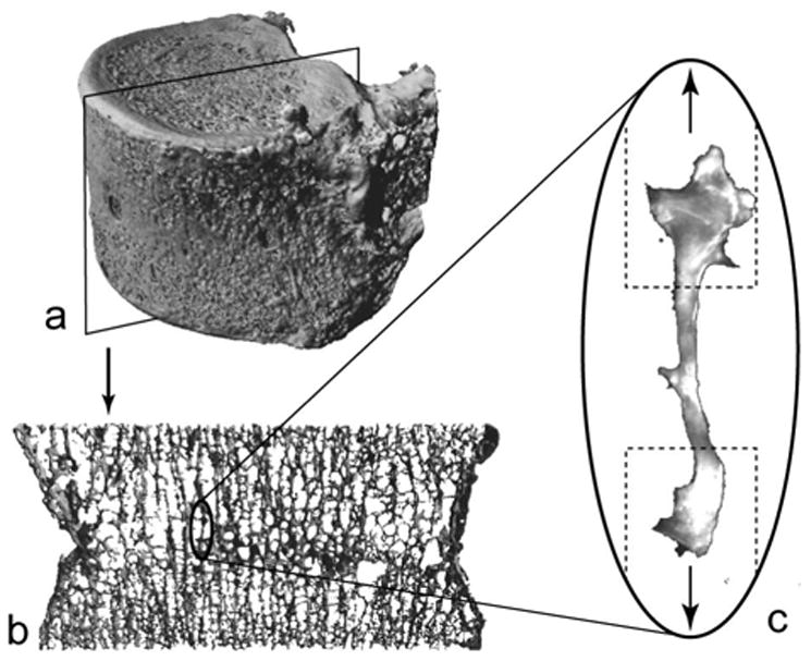
Vertebral bodies (a) were cut into 1 mm thick sagittal sections (b) in order to dissect individual, rod-like, vertical trabeculae (c). The approximate placement of sample holders is indicated with a dashed line and the direction of loading is indicated by arrows.