Figure 1.
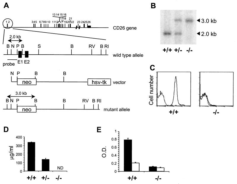
Generation of CD26-deficient mice. (A) Targeting strategy. Exons in the CD26 gene are numbered. The wild-type CD26 allele, targeting construct, and expected mutant allele are shown with exon 1 and 2 of CD26 as filled boxes and the neo and TK genes as open boxes. Restriction sites: B, BglII; N, NcoI; P, PstI; S, SphI; RI, EcoRI, RV, EcoRV. (B) Southern blot containing BglII-digested DNA from CD26 wild-type (+/+), heterozygous (+/−), and homozygous mutant (−/−) mice hybridized with a 0.7-kb BglII–PstI probe specific for regions upstream of the targeting construct. (C) Flow cytometric analysis of CD26 expression at the surface of thymocytes from CD26 wild-type (+/+) and homozygous mutant (−/−) mice stained with anti-CD26 mAb followed by FITC-labeled anti-Ig (solid line) or with FITC-anti-Ig alone (dotted line). (D) Soluble CD26 protein in serum from CD26 wild-type (+/+), heterozygous (+/−), and homozygous mutant (−/−) mice measured by ELISA. ND, not detectable. (E) DPP IV enzyme activity in plasma from CD26 wild-type (+/+) and homozygous mutant (−/−) mice (n = 7). The DPP IV substrate Gly-Pro-pNA (pNA, p-nitroanilide) was incubated in plasma alone (black bars) or plasma containing 10 μM valine-pyrrolidide (open bars).

