
Figure 1.
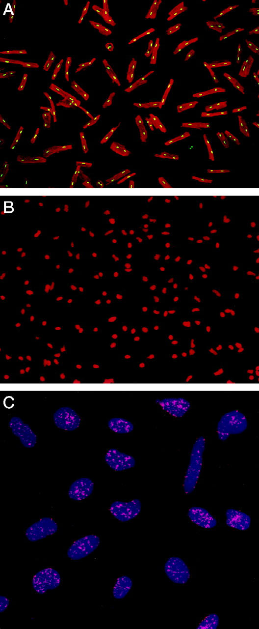
Percoll-treated myocytes (A). Nuclei are stained by PI (yellow fluorescence) and the cytoplasm by α-sarcomeric actin (red fluorescence). Isolated myocyte nuclei are depicted by red fluorescence of PI (B). Nuclei (blue fluorescence) after in situ hybridization with a PNA probe specific for telomeric sequence (C). Red fluorescent dots correspond to individual telomeres. Confocal microscopy; original magnifications, ×150 (A), ×300 (B), ×1500 (C).