
Figure 1.
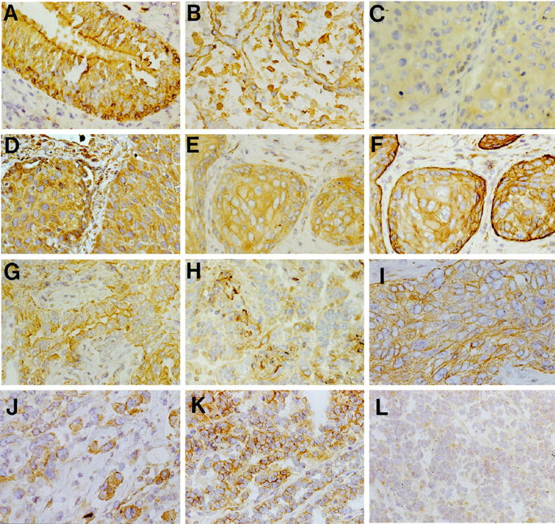
SEMA3F and VEGF levels and localization in normal lung and lung tumors. SEMA3F (A–C, E, G–L) and VEGF (D, F, H) were detected with a secondary biotinylated anti-rabbit antibody. In normal lung (A, B), SEMA3F was detected in all epithelial cells (score 3) with strong membranous staining in bronchial epithelial cells (A) and type II pneumocytes (B). Three examples are given for lung tumors (C–H) for: a squamous cell carcinoma with SEMA3F score 0 to 1 (C) and VEGF score 3 (D); for another squamous cell carcinoma with SEMA3F score 2 (E) and VEGF score 3 (F); and for an adenocarcinoma with SEMA3F score 3 (G) and VEGF score 0 to 1 (H). Note in C and E the diffuse cytoplasmic localization of SEMA3F, and in G the membranous localization of SEMA3F. SEMA3F was located at the basally oriented membrane toward tumor stroma in carcinoma suggesting a function in cellular interactions (I). Cells disseminated in the stroma expressed higher levels of SEMA3F (J) than in lobules. SEMA3F staining was intense and membranous in low grade neuroendocrine carcinoid (K) but diffuse and cytoplasmic in high grade neuroendocrine SCLC tumors (L).