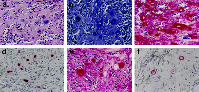
Figure 2.
Analysis of single chondrocytic tumor cells in otherwise matrix-rich small-cell areas. a: Conventional H&E staining. b: Histochemical demonstration of pericellular glycosaminoglycans (toluidine blue). c, e, f: Immunodetection of aggrecan (c), COL2 (e), and COL10 (f) in the extracellular tumor matrix. d: Immunodetection of S-100 protein selectively in the neoplastic chondrocytic cells. Original magnification, ×100.

