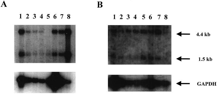
Figure 2.
Northern blot analysis of human tissue for specific expression of hfgl2. A 539-nucleotide long probe from hfgl2 exon I was used. A: lane 1, spleen; lane 2, thymus; lane 3, prostate; lane 4, testis; lane 5, ovary, lane 6, small intestine; lane 7, colon; lane 8, peripheral blood leukocytes. B: lane 1, skeletal muscle; lane 2, uterus; lane 3, colon; lane 4, small intestine; lane 5, bladder; lane 6, heart; lane 7, stomach; lane 8, prostate. Twenty μg of total RNA was added to each lane and hybridized with a human fgl2 cDNA. A glyceraldehyde-3-phosphate dehydrogenase cDNA was used to ensure integrity of the RNA in tissues studied.

