
Figure 5.
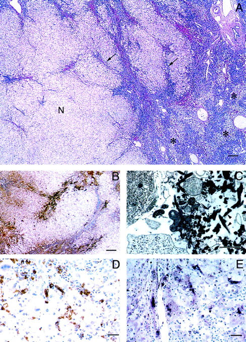
Histopathological and in situ hybridization analysis of fulminant viral hepatitis. A: Surviving nodule (N) of hepatocytes within which are areas of early acute hepatitis with inflammatory cell infiltrates, hepatocellular necrosis, and early collapse (arrows). Remainder of liver showed more advanced total multilobular hepatocellular necrosis (*). B: Fibrin deposits were selectively located in areas of residual parenchyma that showed active, acute necrosis demonstrated by immunoperoxidase using a rabbit-antifibrinogen/fibrin antibody. C: Electron micrograph showing typical early fibrin thrombus comprised of criss-cross fibrin strands (arrowheads) admixed with necrotic cell fragments; the thrombus partially occludes the hepatic sinusoid. Lead citrate stain. D: Immunoperoxidase stain showing that CD68-positive macrophages comprise a significant component of the inflammatory cell infiltrate in areas of bridging necrosis. E: Transcripts of hfgl2 detected in macrophages by in situ hybridization using a 216-nucleotide sequence from the conserved carboxyl end of the gene; compare with D. Digoxigenin-11-UTP-alkaline phosphatase method. Scale bars: A and B, 200 mm; C, 2 mm; D and E, 20 mm.