
Figure 5.
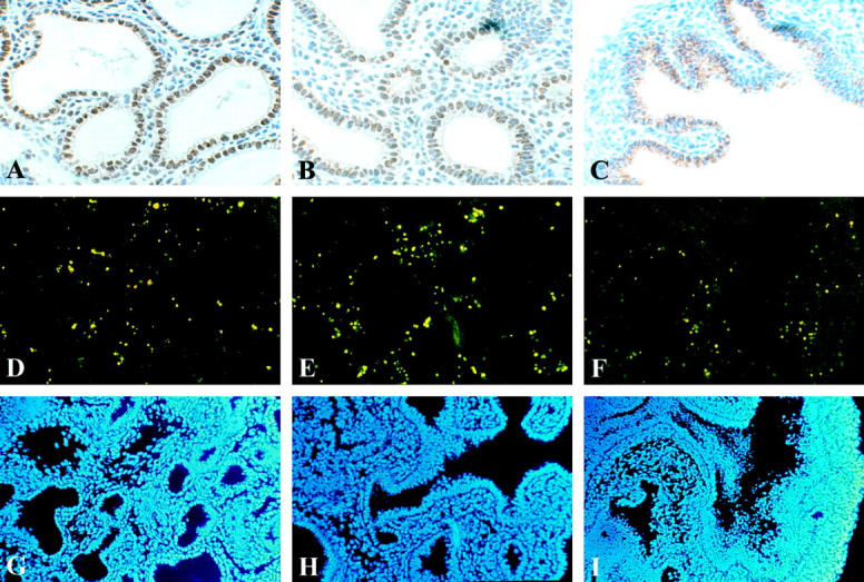
Following a culture period of 4 days, proliferation (A−C) and apoptosis (D−F) were investigated. Immunolocalization of PCNA indicates proliferating cells. Strong PCNA immunoreactivity was observed in the epithelial lining of the terminal buds of the control and DMSO explants, and also, but lower in the mesenchyme (A and B). Surprisingly immunoreactivity was not observed in the mesenchyme and only perinuclear in the explants exposed to Nitrofen (C). Apoptosis in the explants was assessed using TUNEL assay. TUNEL positive cells were observed in all explants in the mesenchyme only, and the number of TUNEL positive cells appeared similar in all explants (D−F). All nuclei were stained using DAPI mounting solution (G−I). All pictures are representative of a series of experiments. All images at same magnification.