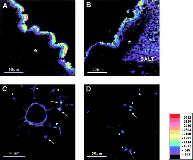
Figure 4.
Immunostaining of CD14, fluorescence. Positive immunostaining is visualized by pseudocolor conversion of the fluorescence image. Bronchial epithelial cells are heavily stained (bronchus of first or second generation A and B), whereas bronchial smooth muscle cells (*, A and B) exhibit no staining for CD14. Cells of the BALT express CD14 (B). Vascular smooth muscle cells of a small partially muscular vessel within the lung parenchyma are positive for CD14 (C). Note that alveolar macrophages (arrows) express CD14 (C and D). In the lung parenchyma, single cells (arrowheads) within the alveolar septum show positive immunostaining for CD14 (D). Scale bar, 50 μm.

