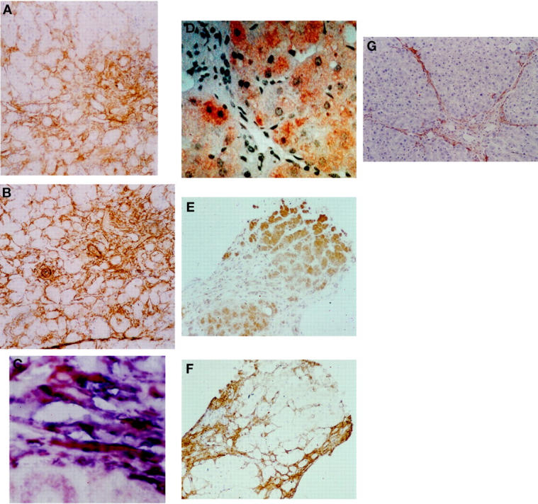
Figure 4.

Cirrhotic liver biopsies were immunostained for p75 as described in Materials and Methods. Clear staining was observed in the areas of fibrosis in a distribution consistent with activated HSC. A representative example of this is demonstrated in A. Analysis of a sequential section for α-SMA confirms that the cells in the fibrotic areas are positive and therefore activated HSC (B; original magnification, ×10). Colocalization for p75 and α-SMA was determined as described in Materials and Methods (C), the majority of the activated HSC in the fibrotic septae are stained by both antibodies resulting in a deep purple color (original magnification, ×40). Staining of cirrhotic biopsies for Fas demonstrates a dramatically different pattern with intense staining of hepatocytes (D; original magnification, ×20) and little or no staining of activated HSC within the fibrotic bands (E), which were demonstrated to be α-SMA-positive (F; original magnification, ×10). Fibrotic rat liver was also stained for p75. Positivity was observed in the myofibroblast-like cells within and adjacent to the fibrotic bands in a distribution consistent with activated HSC (G; original magnification, ×10).
