
Figure 5.
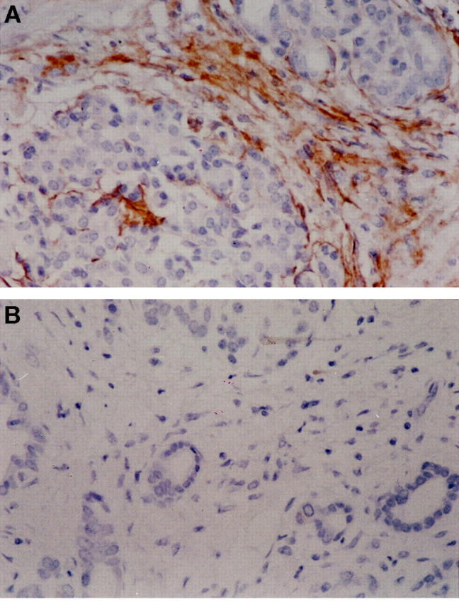
Sections of fibrotic pancreas were immunostained for p75 (A), as described in Materials and Methods. Staining was evident in nerve bundles. In addition, there was clear staining of the fibrotic bands within the pancreas in a distribution entirely consistent with activated PSC and in cells with a myofibroblast-like morphology (original magnification, ×20). A representative negative control for the immunostaining is given in B.