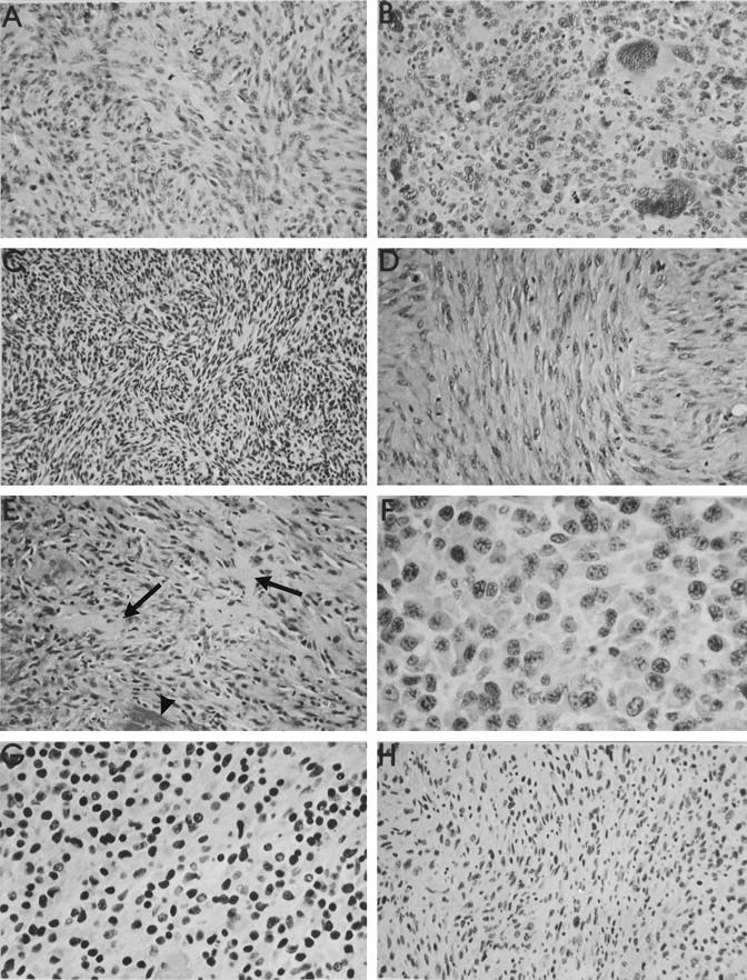
Figure 3.

Representative histological types of soft tissue tumors arising around various subcutaneous implanted biomaterials (all HE stain). A: MFH around a PE implant at 101 weeks, showing a mild storiform pattern and numerous mitotic figures. B: Pleomorphic sarcoma around a PU implant with many large cells with bizarre hyperchromatic nuclei. C: Fibrosarcoma induced by an Al2O3 implant at 103 weeks. Interweaving bundles of small spindle cells are demonstrated. D: High mitotic rate in a leiomyosarcoma around a Ti implant at 107 weeks with typical blunt-ended vesicular nuclei. E: Foci of osteoid matrix (arrows) in an osteosarcoma induced by a PMMA implant after 84 weeks. Foci of calcified matrix were also seen (arrowhead). F: PU-induced epithelioid sarcoma with markedly polygonal tumor cells. G: Small hyperchromatic nuclei in a round cell sarcoma induced by a PU implant after 80 weeks. H: Anisomorphic nuclei in a sarcoma around a Si implant after 90 weeks. No specific morphological pattern identifiable. Hence the classification sarcoma NOS. Objective magnifications, ×40 (F and G) and ×20 (all others).
