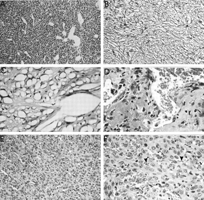
Figure 4.

Histological appearance (HE stain) of sarcomas of vascular origin. A: Malignant hemangiopericytoma around an Al2O3 implant after 46 weeks. Vascular channels of various calibers and shapes are surrounded by relatively small, polygonal cells. Objective magnification, ×10. B: Characteristic low-power (objective magnification, ×10) of a PU-induced hemangiosarcoma, giving a spongy appearance. C: Higher power view (objective magnification, ×40) to demonstrate vessel branching. The numerous tumor vascular spaces are formed by cells with markedly hyperchromatic nuclei. D: Malignant papillary hemangioendothelioma around a NiCr implant at 107 weeks. High-power view (objective magnification, ×40), illustrating two papillary elements, consisting of pleomorphic cells and numerous vascular channels. E: Highly cellular form of an hemangiosarcoma, induced at 90 weeks by a Ti implant. The high degree of vascularity is apparent. Objective magnification, ×20. F: Higher power view of the cellular hemangiosarcoma to demonstrate the marked level of apoptosis (arrowheads). Objective magnification, ×40.
