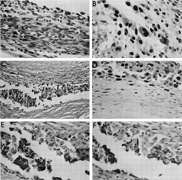
Figure 6.

Immunohistochemical study of preneoplastic lesions. A: Very cellular, preneoplastic lesion in a capsule around a Ti implant. Marked cellular atypia are seen (HE stain). B: PCNA immunohistochemistry of the same lesion as in A. Note the intense nuclear staining in the majority of the atypical cells. C: Preneoplastic lesion in the capsule surrounding a PMMA implant. Despite the relatively thin cellular layer, many cells demonstrate clear atypia (HE stain). D: Marked PCNA staining of the atypical cells in the same case as in C. These cells were immediately adjacent to the implant (upper portion of figure). A gradient of PCNA positivity was observed from the implant toward the peripheral part of the capsule, so that here (lower portion of figure) spindle-shaped fibroblastic cells without atypia showed only faint or no positivity. E: Same case as in C, showing the immunohistochemical reaction with the KiB1R antibody directed against B lymphocytes. A subpopulation of the atypical cells is stained positively. F: Same case as in E, demonstrating a positive subpopulation of atypical cells in the preneoplastic lesion staining for the KiM2R antibody, directed against activated macrophages. Original magnifications, ×20 (C) and ×40 (all others).
