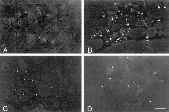
Figure 1.

Fluorescent microscopy of eosinophils in cross-sections of mouse quadriceps muscle. Bright cells are eosinophils. Micrographs are printed so that muscle fibers can be discerned by faint background fluorescence. A: Representative section of 4-week-old C57 quadriceps. One eosinophil is present in the field. Scale bar, 60 μm. B: Representative section of necrotic region of 4-week-old mdx quadriceps. Scale bar, 50 μm. C: Representative section of a regenerated region of 30-week-old mdx quadriceps. Scale bar, 50 μm. D: Representative section of quadriceps from a 4-week-old perforin-deficient, mdx mouse. Scale bar, 60 μm.
